1
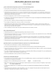
Med Oral 2003;8:10-18
Mucositis en hemopatías malignas
Mucositis in hematological malignancies
Protocolo de estudio y tratamiento de la Mucositis bucal
en los pacientes con hemopatías malignas
Margarita Puyal Casado (1), Carmen Jiménez Martínez (2), Eduardo Chimenos Küstner
(1,3), José López López (1,4), Antonio Juliá ( 2)
(1)
(2)
(3)
(4)
Médico especialista en Estomatología.
Servicio de Hematología Clínica. Hospital General Vall d’Hebrón.
Profesor titular de Medicina Bucal. Universidad de Barcelona.
Profesor asociado de Medicina Bucal. Universidad de Barcelona.
Correspondencia:
Margarita Puyal Casado
Paseo Maragall, 298 enlo 3ª
08031 Barcelona
Tel. 933572488
E-mail: [email protected]
Recibido: 26-8-2001 Aceptado: 12-10-2002
Puyal-Casado M, Jiménez-Martínez C, Chimenos-Küstner E, López-López J, Juliá A.
Protocolo de estudio y tratamiento de la Mucositis bucal en los pacientes con hemopatías malignas. Med Oral 2003;8:10-18
© Medicina Oral S. L. C.I.F. B 96689336 - ISSN 1137 - 2834
RESUMEN
Palabras clave: Hemopatía maligna, Quimioterapia,
Radioterapia, Mucositis.
Objetivo: Conocer la incidencia real de las lesiones en
la cavidad bucal de los enfermos con hemopatías malignas, las debidas a la enfermedad y las ocasionadas
por los tratamientos recibidos, con el fin de prevenir,
diagnosticar y tratar las manifestaciones orales de la
hemopatía así como las mucositis originadas por los
diferentes tratamientos de quimioterapia o radioterapia.
Diseño: se revisaron a pacientes con hemopatías malignas en fase de tratamiento o a los que se les iba a
realizar un transplante hematopoyético. Se les realizó
un estudio clínico, y en casos necesarios un cultivo
microbiológico o un estudio anatomopatológico.
Resultados: la realización de un protocolo en estos
pacientes para el control de sus lesiones orales así
como la realización de una pauta profiláctica y de tratamiento para sus lesiones.
Conclusión: mejor conocimiento y manejo de los pacientes con hemopatías malignas.
1.- INTRODUCCION
La boca es una cavidad donde con frecuencia se producen lesiones durante el curso de las hemopatías.
Estas lesiones pueden ser bien una manifestación primaria de la enfermedad o una secundaria a los diferentes tratamientos a que son sometidos estos pacientes.
Las lesiones primarias o específicas en la boca están
presentes en la mitad de los pacientes con leucemia
aguda (LA), sobre todo en las linfoblásticas (LAL),
algo menos en las mieloides y dentro de este grupo
destaca la infiltración gingival en las mielomonocíticas
(LAM5 de la clasificación FAB). Una hiperplasia
gingival que aparece sin motivo evidente
farmacológico o periodontal puede ser el primer síntoma de una leucemia e incluso preceder a otras manifestaciones(1-3). En la tabla 1 se exponen las prin10
Med Oral 2003;8:10-18
Mucositis en hemopatías malignas
Mucositis in hematological malignancies
cipales manifestaciones bucales de las hemopatías malignas.
La presentación de un linfoma primario en la cavidad
oral es rara (1,2), excepto en los pacientes infectados
por el virus de la inmunodeficiencia humana. Los
linfomas no hodgkinianos (LNH) son los que con mayor frecuencia se ubican en esta región. El anillo de
Waldeyer está infiltrado en la mitad de estos casos y
menos a menudo lo están también la encía, la lengua
o el paladar. Por otro lado, la lesiones de la cavidad
oral en los pacientes con mieloma múltiple son poco
frecuentes y suelen ser lesiones osteolíticas en los
maxilares que ocasionalmente se presentan como fracturas patológicas, parestesias, disfunción de la articulación temporomandibular (ATM) y tumoraciones de
los tejidos blandos de la boca (3-15).
Las diferentes pautas de tratamiento que reciben estos
pacientes con citostáticos, radioterápicos y antibióticos
pueden ocasionar la aparición de diversas lesiones
secundarias en la cavidad oral (tabla 2), entre las cuales la más importante es la mucositis. Las alteraciones
de la mucosa bucal deberían denominarse estomatitis,
ya que el término mucositis es muy inespecífico y hace
referencia a cualquier membrana mucosa del organismo. Sin embargo en la literatura internacional parece
haber un acuerdo en el uso de la palabra mucositis
oral (o bucal) para indicar las alteraciones que los tratamientos oncológicos producen en la mucosa bucal.
Muchos fármacos provocan mucositis (tabla 3). Los
citostáticos (6) tienen una acción tóxica directa al destruir la membrana basal del epitelio y el parénquima
de las glándulas salivales y otra indirecta derivada de
la mielosupresión con sus manifestaciones
hemorrágicas e infecciosas (2). Algunos pacientes también experimentan los efectos adversos de la radioterapia. Las lesiones que provocan las radiaciones
ionizantes en la boca y las glándulas salivales están
caracterizadas tanto por su cronología como por el
efecto dosis. Las úlceras y la hiposialia que se observan entre los 8-15 días del inicio de la radioterapia se
consideran como lesiones inmediatas y consecuencia
directa de su acción que es reversible entre los 15-21
días postratamiento. Las caries que se presentan a medio plazo entre los 3 y 6 meses de finalizada la radioterapia sobre las diferentes estructuras son debidas a
la hiposialia generada por la sialoadenitis y el trismo
está provocado por la fibrosis de los músculos
masticadores. Respecto al efecto dosis, 10 Gy provocan una hiposialia que resulta reversible hasta dosis
de 50 Gy; una dosis acumulada de 60 Gy induce lesiones irreversibles por la atrofia y necrosis de los
acinos serosos de las glándulas salivares junto con la
atrofia parcial de las glándulas mucosas y mixtas. La
saliva de estos pacientes es más espesa por el incremento de las concentraciones iónicas de Na+, Cl-,
Ca++, Mg++ y de las proteínas. Las tasas de irradiación superiores a los 5 Gy a largo plazo, pueden producir a partir de los 5-6 meses de haber recibido el
tratamiento una disminución del aporte sanguíneo al
hueso con necrosis y una reparación ósea anormal.
Ejemplos son la amplia gama de malformaciones dentarias y/ o agenesias en los maxilares que desarrollan
los muy jóvenes con dientes en estadio preeruptivo o
erupcionados, pero inmaduros (16-19).
La incidencia de las lesiones bucales varía según la
hemopatía, el tipo de tratamiento aplicado (20) y el
estado bucal previo a la aparición de la enfermedad.
Sin embargo, la acumulación de factores hace que el
85 % de los pacientes sometidos a tratamiento con
dosis elevadas desarrollen complicaciones en la cavidad oral.
2. - OBJETIVOS DEL ESTUDIO
Es realizar un protocolo que ayude a:
·Prevenir la aparición de mucositis en los pacientes
con hemopatías malignas ingresados.
·Diagnosticar y tratar correctamente las lesiones en la
cavidad oral.
·Conocer la incidencia real de las lesiones primarias y
secundarias en la cavidad bucal.
·Controlar la aparición de posibles cepas bacterianas
resistentes a los fármacos de uso habitual.
3. - MATERIAL Y METODOS
CRITERIOS DE INCLUSIÓN
Se revisaron los pacientes con hemopatías malignas
ingresados en la Unidad de Hematología Clínica susceptibles de presentar lesiones en la cavidad bucal, ya
específicas por la propia enfermedad de base o secundarias a los tratamientos que reciben con antibióticos,
citostáticos y radiaciones. Se incluirán los pacientes
pertenecientes a los siguientes grupos de riesgo:
·Pacientes con hemopatías malignas y/ o neutropenia
grave en fase de tratamiento.
·Pacientes a los que se va realizar un trasplante
hematopoyético.
4.- MUCOSITIS
Como ya se ha indicado anteriormente, en la literatura
internacional parece haber un acuerdo en el uso de la
palabra mucositis oral (o bucal) para indicar las alteraciones que los tratamientos oncológicos producen
11
Med Oral 2003;8:10-18
Mucositis en hemopatías malignas
Mucositis in hematological malignancies
en la mucosa bucal, aunque las lesiones de la mucosa
bucal deberían denominarse estomatitis, ya que el término mucositis es muy inespecífico y hace referencia
a cualquier membrana mucosa del organismo.
4.1-Factores predisponentes para la mucositis
El estado de salud de la cavidad oral previo a la enfermedad es uno de los principales factores
condicionantes del desarrollo del grado de la mucositis
(21), de la duración y de su evolución. Otros factores
que modulan la sintomatología de la mucositis son las
hemopatías con manifestación bucal, los citostáticos,
la radioterapia, así como el estado de inmunodepresión
o la coexistencia de otras enfermedades sistémicas
como la diabetes, la insuficiencia renal o los trasplantes de órganos (tablas 1 y 2), así como una correcta
higiene bucal (21).
LEUCEMIAS
Palidez de la mucosa
Infiltrados gingivales
Hemorragias gingivales
Petequias
Úlceras de la mucosa
necróticas
hemorrágicas
LINFOMAS
MIELOMAS
No-hodgkinianos
Anillo de Waldeyer
Encía Lengua Paladar
Primarios en el SIDA
Lesiones osteolíticas
maxilares
Fracturas patológicas
Parestesias
Disfunción ATM
Tumoraciones de los
tejidos blandos bucales
0.- Normalidad
I.- Eritema generalizado: mucosa rosada no dolorosa
y con abundante saliva. Voz normal.
II.- Eritema, úlceras poco extensas, se mantiene la deglución de sólidos.
III.- Úlceras extensas, encías edematosas, saliva espesa; se mantiene la capacidad de deglutir líquidos. Dolor. Dificultad para hablar.
IV.- Úlceras muy extensas, encías sangrantes, infecciones, no hay saliva, es imposible deglutir. Dolor muy
intenso.
5.- METODOLOGIA DIAGNOSTICA
Al ingreso del paciente en la Unidad de Hematología
Clínica, un estomatólogo o un odontólogo deberá explorarlo y diagnosticar el estado de salud bucal basal,
cumplimentará la ficha dental y realizará el seguimiento de la mucositis (ver figura 1) (24).
En las lesiones en las que por la clínica no pueda filiarse
el diagnóstico se realizarán los siguientes estudios complementarios:
5.1- Microbiología
De los exudados de la cavidad oral: se tomarán las
muestras de lugar de las lesiones ya sean en la lengua,
la mucosa yugal o el surco gingival y de los frotis se
realizarán cultivos microbiológicos para gérmenes
aerobios, anaerobios y hongos
5.2- Anatomía patológica
De los nódulos, los agrandamientos gingivales o de
los infiltrados: se tomarán muestras para el estudio
histológico.
Tabla 1. Manifestaciones bucales de las hemopatías malignas
FÁRMACOS
-Mucositis
-Xerostomías
-Infecciones
-Diátesis hemorrágica
RADIACIONES IONIZANTES
6.- TRATAMIENTO DE LA MUCOSITIS
(Tabla 4)
La indicación se hace en función del grado de
mucositis y se instaura el correspondiente a un grado
por encima al que se ha establecido en el diagnóstico.
Grados 0 y I
El adiestramiento del paciente en las siguientes medidas de higiene:
1. El correcto uso de un cepillo dental suave después
de las comidas.
2. El colutorio de clorhexidina en enjuagues después
del cepillado.
3. La ingesta elevada de líquidos para mantener la salivación.
4. La integridad de los labios se mantiene con la aplicación tópica de vaselina.
Grado II
Las medidas de tratamiento de los grados 0 y I se aplicarán ahora cada 4 horas y se añaden el agua
-Inmediatas Mucositis Disgeusia
Glosodinia Xerostomía Disfagia
Trismo
-A medio plazo Caries Necrosis de
la mucosa
-A largo plazo Osteorradionecrosis
Alteraciones del desarrollo del
germen dental Agenesias
Alteraciones coronales Alteraciones
radiculares
Tabla 2. Manifestaciones por los tratamientos en las hemopatías malignas
4.2- Grados de mucositis
En 1979, la O.M.S. (4,22,23) definió el grado de las
lesiones de la mucositis según la severidad de las mismas. Se establecen así cinco grados, del 0 al IV, siendo el 0 la ausencia de lesiones y el IV la presencia de
lesiones graves en extensión y profundidad.
12
Med Oral 2003;8:10-18
CITOSTÁTICOS
Metotrexato
Procarbacina
Tioguanina
Mercaptopurina
Citarabina
Fluoruracilo
Floxuridina
Vinblastina
Vincristina
Dactinomicina
Daunorrubicina
Doxorrubicina
Mitramicina
Bleomicina
Asparaginasa
Mucositis en hemopatías malignas
Mucositis in hematological malignancies
INMUNOSUPRESORES
Corticoides
Azatioprina
Ciclosporina A
OTROS FÁRMACOS
Fármacos
xerostomizantes
Ansiolíticos
Antidepresivos
Antihistamínicos
Estimulantes
simpaticomiméticos
Antiparkinsonianos
Antipsicóticos
Agrandamiento gingival
Hidantoínas
Ciclosporina A
Antibióticos de amplio
espectro
Grado/
curas
O
I
II
III
Examen
cada
24 h
24 h
12 h
8h
Curas
cada
Después
de las
comidas
6h
4h
2h
2h
Enjuagues
CHX /
H2O
CHX /
H2O / SB
CHX /
H2O / SB
CHX / SB
/
Antifúngicos
CHX / SB
/
Antifúngicos / *
Lubricar
labios
Cada 8h
Cada 6 h
Cada 4 h
Cada 2 h
Cada hora
IV
4h
Tabla 4. Grado y tratamiento de las mucositis
CHX: Digluconato de clorhexidina al 0,12 % SB: Solución de bicarbonato 7,5 cc en 500 cc H2O o bien 1,4 g, en 100 ml de agua destilada). H2O:
Agua * Prescripción específica según el estado del paciente Otras
recomendaciones: Antifúngicos orales: nistatina en enjuagues. Mantenerlo durante 4 minutos en la boca y tragarlo o escupirlo. Evitar los
alimentos ácidos, muy calientes o muy fríos y con superficies duras.
Ingesta elevada de líquidos (> de 2 l/ 24 h) Gel de CHX en aplicación
tópica sobre las lesiones de la mucosa. Cepillo blando: Vitis ultra suave®
Cepillo muy blando: Vitis cirugía®
Tabla 3. Fármacos que producen mucositis
bicarbonatada y nistatina (25-27) en solución, 5 cc
cada 6 horas, como colutorios. Las prótesis removibles
se mantendrán en la boca solo durante las comidas.
Grados III y IV
Las medidas de tratamiento de la mucositis de grado
II se complementan con los tratamientos tópicos y
sistémicos según la etiología de las lesiones.
Las infecciones micóticas de la cavidad oral se tratarán con: nistatina en solución 5 cc cada 4 horas y
fluconazol (25-27) en solución 200 mg al día, por vía
oral.
Las infecciones herpéticas locales se tratan con
aciclovir en crema 5 aplicaciones al día.
Las úlceras se tratan localmente con 4 o 6 aplicaciones diarias de cualquiera de las siguientes fórmulas
magistrales:
· Acetónido de fluocinolona al 0,1% en orabase.
· Acetónido de triamcinolona al 0,1% en orabase.
· Hidrocortisona al 1% en orabase.
El hematólogo indicará el tratamiento sistémico en estos pacientes para evitar la extensión de las infecciones locales y aliviar el dolor.
instruirá al paciente en las medidas de higiene oral.
«Primum non nocere» o evitar los irritantes locales de
tipo físico, químico o térmico. Para ello es preciso seguir las siguientes pautas de profilaxis y diagnóstico.
1.- La retirada de las prótesis removibles que actúan
como reservorios de gérmenes, en especial de Candida
sp, mientras dura la fase aguda de la mucositis y la
higiene con productos liberadores de cloro o bien con
clorhexidina.
2.- La valoración de los tratamientos ortodóncicos tanto los fijos como los removibles y el efecto irritante
que pueden tener sobre la cavidad bucal del enfermo.
Consultar si procede con el ortodoncista del paciente.
3.- Uso de colutorios sin alcohol:
· La clorhexidina en forma de digluconato (es un
antibacteriano de amplio espectro que también es activo frente a Candida sp). Se emplea a concentraciones de 0,12 % y de 0,2 %. La concentración de 0,12 %
tiene presentación como colutorio y la de 0,2 % en
forma de gel bioadhesivo (29-31).
· El agua bicarbonatada alcaliniza el pH de la saliva
dificultando el crecimiento de Candida sp y la descalcificación del esmalte grabado por los ácidos.
· El agua oxigenada diluida a partes iguales en agua
arrastra los restos alimenticios y los detritos que se
acumulan sobre los dientes y la mucosa.
4.- Los alimentos de consistencia blanda y a temperatura ambiente son los indicados para evitar el trauma
7. - MEDIDAS DE PROFILAXIS DE LA
MUCOSITIS
Los pacientes que mantienen una higiene cuidadosa
de la boca presentan una mucositis de menor gravedad y duración que aquellos que no la realizan (21,
28). Si bien la mucositis mejora al resolverse la
neutropenia del paciente, la prevención con la identificación y el tratamiento precoz de las lesiones
bucodentales revisten una importancia fundamental en
la calidad de vida de estos enfermos. La enfermera
13
Med Oral 2003;8:10-18
Mucositis en hemopatías malignas
Mucositis in hematological malignancies
Diagnóstico del estado de salud bucal
No lesiones bucales
PROFILAXIS
MUCOSITIS
Lesiones de etiología dudosa
Microbiología
(exudados)
Tratamientos
Tópico
Sistémico
Mucositis de la cavidad oral
Valoración de la gravedad
Anatomía Patológica
(biopsia)
TRATAMIENTO DE LA MUCOSITIS
Lesiones
Específicas o primarias
Lesiones
Secundarias o inespecíficas
Tratamiento
Tratamiento
Tópico
Sistémico
Fig. 1. Lesiones en la cavidad oral de los pacientes con hemopatías
malignas: diagnóstico, profilaxis y tratamiento
térmico o por el roce. Las dietas bajas en hidratos de
carbono son aconsejables para prevenir las caries.
5.-La ingesta elevada de líquidos mejora la hiposialia.
6.- La higiene meticulosa de la dentadura y las encías
siempre que el recuento de plaquetas sea superior a
5x10⁹/l, con un cepillo dental muy suave Vitis cirugía®. No se usará dentífrico desde el grado I de
mucositis. Los dentríficos fluorados y el cepillado con
cepillo suave Vitis Ultrasuave ® están permitidos en
los períodos sin mucositis. (32).
transplantation, with clinical assessment and microbiological
culture or histological study where required.
A protocol is developed in these patients for the prophylaxis,
control and management of their oral lesions, thus contributing
to improve the knowledge and treatment of patients with
hematological malignancies.
Key words: Malignant hematological disease, chemotherapy,
radiotherapy, mucositis.
1.- INTRODUCTION
Oral lesions are often observed in the course of hematological
disorders. As such they may constitute primary manifestations
of the disease or represent alterations secondary to the
treatments provided. In this context, primary or specific oral
lesions are present in half of all patients with acute leukemia
(AL), particularly in the lymphoblastic variants of the disease
(ALL). Such oral alterations are somewhat less common in the
case of acute myeloid leukemias, and within this group of
disorders special mention may be made of the gingival
infiltrations characterizing acute myelomonocytic leukemia
(AML5 of the FAB classification). Gingival hyperplasia
developing with no apparent pharmacological or periodontal
cause may be among the first signs of leukemia and can even
precede other manifestations (1-3). Table I describes the principal oral manifestations of hematological malignant processes.
The development of a primary lymphoma in the oral cavity is
rare (1,2), except in patients infected with the human
immunodeficiency virus (HIV). Non-Hodgkin lymphomas
(NHL) are the presentations most often found in this region.
Waldeyer’s ring is seen to be infiltrated in half of these cases,
while associated involvement of the gums, tongue or palate is
less common. On the other hand, the oral lesions found in
patients with multiple myeloma are infrequent and tend to
comprise maxillary osteolytic lesions which occasionally
manifest as pathological fractures, paresthesias,
temporomandibular joint (TMJ) dysfunction and tumors of the
oral soft tissues (3-15).
ENGLISH
A protocol for the evaluation and treatment of oral
mucositis in patients with
hematological malignancies
PUYAL-CASADO M, JIMÉNEZ-MARTÍNEZ C, CHIMENOS-KÜSTNER
E, LÓPEZ-LÓPEZ J, JULIÁ A. A PROTOCOL FOR THE EVALUATION AND TREATMENT OF ORAL MUCOSITIS IN PATIENTS WITH
HEMATOLOGICAL MALIGNANCIES.
MED ORAL 2003;8:10-18
SUMMARY
A study is made to determine the true incidence of oral lesions
attributable to hematological malignancies and their therapies,
with the purpose of preventing, diagnosing and treating the
oral manifestations of the hematological disorder and the
situations of mucositis produced by the different chemo or
radiotherapeutic regimens administered.
A review was made of patients with hematological malignancies
undergoing treatment or programmed for hematopoietic
14
Med Oral 2003;8:10-18
Mucositis en hemopatías malignas
Mucositis in hematological malignancies
3.- MATERIAL AND METHODS
The different therapeutic regimens used in such patients,
involving cytostatic drugs, radiotherapy and antibiotics, can
lead to the development of secondary oral lesions (Table 2), of
which mucositis is the most important representative. In this
sense, alterations of the oral mucosa should be referred to as
stomatitis, since the term «mucositis» is very nonspecific and
in fact refers to any mucosal membrane in the body. However,
there appears to be consensus in the international literature
over the use of the term «oral (or buccal) mucositis» in reference
to the oral mucosal alterations caused by the different
oncological treatments.
Many drugs are able to produce mucositis (Table 3). In this
context, cytostatic agents (4) exert direct toxic action by
destroying the epithelial basal membrane and salivary gland
parenchyma, and moreover exert indirect action as a result of
myelosuppression with its hemorrhagic and infectious
manifestations (2). Some patients also suffer adverse effects as
a result of radiotherapy. In this sense, the oral and salivary
gland lesions caused by ionizing radiations are characterized
by their time-sequence and dose dependency. The ulcers and
diminished salivary flow (hyposialosis) seen between 8-15 days
after the start of radiotherapy are regarded as immediate and
direct lesions that prove reversible 15-21 days after treatment.
The caries seen at middle term 3-6 months after the end of
radiotherapy are in turn attributed to the diminished salivary
flow caused by the radiation-induced sialoadenitis, while
trismus develops as a consequence of fibrosis of the masticatory
muscles. Regarding the dose-dependency of the observed
lesions, 10 Gy induces hyposialosis which proves reversible
up to a dose of 50 Gy. In turn, an accumulated dose of 60 Gy
induces irreversible lesions as a result of atrophy and necrosis
of the serous acini of the salivary glands, together with partial
atrophy of the mucosal and mixed glands. The saliva of these
patients is thicker as a result of increments in the concentrations
of different ions (Na+, Cl-, Ca2+, Mg2+) and proteins. Irradiation
doses of over 5 Gy over the long term (i.e., starting 5-6 months
after treatment) can reduce blood flow to the bone, thus giving
rise to bone necrosis and abnormal bone repair. Examples of
these phenomena include a broad range of dental malformations
and/or ageneses in the jaws of very young patients with preeruptive or erupted but immature teeth (16-19). The incidence
of oral lesions varies according to the blood disorder involved,
the type of treatment provided (20), and the oral condition
prior to development of the disease. However, the accumulation
of contributory factors causes 85% of patient subjected to highdose treatments to develop oral complications.
Inclusion criteria. A review was made of the patients with
hematological malignancies admitted to the Clinical
Hematology Unit and susceptible to develop oral cavity lesions
– either specific of the background disease (i.e., primary) or
secondary to treatment received in the form of antibiotics,
cytostatic drugs and radiotherapy. Patients belonging to the
following risk groups were included: (a) malignant
hematological disease and/or severe neutropenia subjected to
treatment; (b) patients programmed for hematopoietic
transplantation.
LEUKEMIAS
Pale mucosa.
Gingival infiltrates.
Gingival bleeding.
Petechiae.
Mucosal ulcerations:
*necrotic.
*hemorrhagic
LYMPHOMAS
(non-Hodgkin).
Infiltrates:.
gingival.
lingual.
palatal.
Primary in AIDS
MYELOMAS
Maxillary osteolysis.
Pathological fractures.
Paresthesias.
TMJ dysfunction.
Oral soft tissue tumors
Table 1. Oral manifestations of malignant hematological disorders.
INDUCED BY IONIZING
RADIATIONS
DRUG-INDUCED
Mucositis.
Xerostomia.
Infections.
Hemorrhagic diathesis
-Immediate.
Mucositis.Dysgeusia.Glossodynia.Xerostomia.Dysphagia.Trismus.
-Middle-term.
Caries.
Mucosal necrosis.
-Long-term.
Osteoradionecrosis.Dental germinal
development
alterations.Ageneses.Coronal
alterations.Root alterations.
Table 2. Oral manifestations of treatments for malignant hematological
disorders.
CYTOSTATICS
Methotrexate.Procarbazine.Thioguanine.Mercaptopurine.Cytarabine.Fluorouracil.Floxuridine.Vinblastine.Vincristine.Dactinomycin.Daunorrubicin.Doxorubicin.Mitramycin.BleomycinAsparaginase
2.- STUDY OBJECTIVES
The aim of the present study was to develop a protocol capable
of contributing to: (a) prevent the appearance of mucositis in
hospitalized patients with malignant hematological disease;
(b) diagnose and correctly treat the lesions of the oral cavity;
(c) determine the true incidence of primary and secondary
lesions of the oral cavity; and (d) control the possible
appearance of bacterial strains resistant to the habitually used
drug substances.
IMMUNOSUPPRESSORS
Corticoids.
Azathioprine.
Cyclosporine A.
Table 3. Drugs which induce mucositis
15
OTHER DRUGS
Xerostomia.
Anxiolytic agents.
Antidepressants.
Antihistamines.
Sympathomimetic
stimulants.
Antiparkinson
medication.
Antipsychotics.
Gingival enlargement.
-Hydantoin.
-Cyclosporine A.
Disappearance of
saprophytic flora.Broad
spectrum antibiotics
Med Oral 2003;8:10-18
Mucositis en hemopatías malignas
Mucositis in hematological malignancies
4.- MUCOSITIS
Grades 0 and I
The patients should be instructed on the following hygiene
measures: (a) correct and gentle tooth brushing after meals; (b)
chlorhexidine mouth rinsing after brushing; (c) fluid intake to
maintain salivation; and (d) preservation of lip integrity by
applying topical vaseline.
Grade II
The treatment measures defined for mucositis grade 0 and I are
to be applied every four hours, adding carbonated water and
nystatin (25-27) solution (5 ml every 6 hours) as rinses.
Removable dentures are to be kept in the mouth only during
mealtimes.
Grades III and IV
The treatment measures defined for mucositis grade II are to be
complemented with the topical and systemic treatments
indicated according to the etiology of the lesions. Fungal
infections of the oral cavity are to be treated with nystatin
solution (5 ml every four hours) and fluconazole (25-27) in
solution (200 mg/day via the oral route). In turn, local herpetic
infections should be treated with aciclovir cream (5 applications
a day). Lastly, ulcerations are to be treated locally with 4-6
daily applications of any of the following magistral
formulations: (a) 0.1% fluocinolone acetonide in orabase; (b)
0.1% triamcinolone acetonide in orabase; or (c) 1%
hydrocortisone in orabase.
The hematologist should in turn prescribe the systemic therapy
required to avoid the spread of local infectious processes and
for providing pain relief.
As has been commented above, there appears to be consensus
in the international literature over use of the term «oral (or
buccal) mucositis» in reference to the oral mucosal alterations
caused by oncological treatments - though buccal mucosal
lesions should actually be referred to as «stomatitis», since
mucositis is nonspecific and in fact refers to any mucosal
membrane in the body.
4.1.- Factors predisposing to mucositis
The health condition of the oral cavity prior to development of
the disease is one of the conditioning factors of mucositis (21),
its duration and course. Other factors which modulate the
symptomatology of mucositis include hematological disorders
with oral manifestations, cytostatics, radiotherapy, and the
existence of immune depression or other concomitant systemic
illnesses such as diabetes, renal failure or organ transplantation
(Tables 1 and 2), as well as correct oral hygiene (21).
4.2.- Grades of mucositis
In 1979, the World Health Organization (WHO)(4,22,23)
defined the degree of mucositis according to the severity of the
lesions. Five grades were established (from 0 to IV), grade 0
representing the absence of lesions and grade IV the presence
of severe lesions in terms of extent and depth: 0 = normality; I
= generalized erythema (painless pink mucosa with abundant
saliva and normal voice function); II = erythema involving
small ulcerations and preserved solid swallowing capacity; III
= extensive ulcers with edematous gingival tissue and thick
saliva, preserved liquid swallowing capacity, pain and speech
difficulties; IV = very extensive ulcers with bleeding gums,
infections, the absence of saliva, incapacity to swallow, and
intense pain.
Grade
5.- DIAGNOSTIC METHODOLOGY
Upon patient admission to the Clinical Hematology Unit, the
stomatologist or dentist should conduct an exploration and
diagnosis of the basal oral health status, complemented by the
dental record and follow-up of mucositis (Fig. 1)(24).
In those cases involving lesions which cannot be classified,
the diagnosis should be based on the following supplementary
studies:
5.1.- Microbiology
Oral cavity exudate samples should be collected from the lesion
sites involving the tongue, jugal mucosa or gingival sulcus.
The resulting smears should be subjected to microbiological
culture for the isolation of aerobes, anaerobes and fungal
species.
5.2.- Histopathology
Samples are to be collected from any possible nodules, gingival
enlargement or infiltrates for histological study.
O
I
II
II
III
IV
IV
Examination every
24 h
24 h
12 h
8h
4h
Application every
After
meals
6h
4h
2h
2h
Mouthrinses
CHX /
H2O
CHX /
H2O / SB
CHX /
H2O / SB
CHX / SB
/Antifungals
CHX / SB
/Antifungals / *
Every 8 h
Every 6 h
Every 4 h
Every 2 h
Every
hour
Respect
sleep..Leave
dentures.
.Normal
brush and
toothpaste
Leave
dentures
for social
life..Soft
brush.Vitis
Ultrasuave®
Dentures
only for
meals..Very soft
brush.Vitis
Cirugía®
Remove
dentures..Very soft
brush.Vitis
Cirugía®
No
dentures.Brushing
depends
on
complete
blood
count.Vitis
Cirugía®
Lubricate
lips
Others
Table 4. Mucositis grades and treatment.
CHX: 0.12% chlorhexidine digluconate.SB: Solution of bicarbonate (7.5
ml in 500 ml of H2O or alternatively 1.4 g in 100 ml of distilled water).H2O:
water.* Specific prescription according to patient condition .Other
recommendations:.Oral antifungals: nystatin mouthrinse. Keep for 4
minutes in mouth and either swallow or discard.Avoid acid, very hot or
very cold foods and hard surfaces.Important fluid intake (> 2 liters
daily)..CHX gel for topical application to mucosal lesions.Soft toothbrush:
Vitis Ultrasuave®.Very soft toothbrush: Vitis Cirugía®..
6.- TREATMENT OF MUCOSITIS
(Table 4)
The treatment indications are based on the grade of mucositis,
and therapy corresponding to one grade higher than that
established at initial diagnosis is to be provided.
16
Med Oral 2003;8:10-18
Mucositis en hemopatías malignas
Mucositis in hematological malignancies
Diagnosis of oral health status
Lesions of uncertain etiology
No oral lesions
PROPHYLAXIS
MUCOSITIS
Microbiology
(exudates)
Treatments:
Topical
Systemic
Oral cavity mucositis
Severity assessment
Histopathology
(biopsy)
Specific or primary
lesions
Treatment
TREATMENT OF MUCOSITIS
Secondary or nonspecific
lesions
Treatments:
Topical
Systemic
Fig. 1. Oral cavity lesions in patients with malignant hematological
disease: diagnosis, prophylaxis and treatment
7.- MUCOSITIS PROPHYLACTIC
MEASURES
(f) Careful dental and gingival hygiene is indicated provided
the platelet count is over 5 x 10⁹ platelets/l, using a very soft
tooth brush (Vitis Cirugía®). Toothpaste should be avoided
above mucositis grade I. Fluorated toothpastes and brushing
with a soft brush (Vitis Ultrasuave®) is acceptable in periods
without mucositis (32).
Patients who observe careful oral hygiene suffer less severe
mucositis, and its duration is moreover shorter than in patients
who neglect such hygiene (21,28). Although mucositis
improves upon resolving patient neutropenia, prevention and
the identification and early treatment of buccodental lesions is
of paramount importance for ensuring adequate patient quality
of life. The nursing personnel should instruct the patient on
correct oral hygiene. In this context, the influence of local
irritants (physical, chemical or thermal) should also be avoided.
The following prophylactic and diagnostic steps are indicated
to this effect:
(a) Avoidance of removable dentures which serve as reservoirs
for microorganisms, especially Candida spp., for the duration
of the acute phase of mucositis. Measures of hygiene involving
chlorine-releasing products or chlorhexidine also apply here.
(b) Assessment of orthodontic treatment (both fixed and
removable) and the irritating actions they may exert upon the
oral cavity of the patient. The patient orthodontist should be
consulted to this effect.
(c) Mouthrinses without alcohol.
Chlorhexidine digluconate (a broad spectrum antibacterial
agent which is also active against Candida spp.) can be applied
in solution form at concentrations of 0.12% and 0.2%.. The
lesser concentration (0.12%) is supplied as a mouthrinse, while
the 0.2% presentation is used as a bioadhesive gel (29-31).
Carbonated water ensures an alkaline salivary pH, which
contributes to counter the growth of Candida spp. and acidetched enamel decalcification.
Hydrogen peroxide diluted in equal proportions of water helps
clear food remains and detritus that accumulate on the teeth
and mucosas.
(d) Soft foods at room temperature are indicated for avoiding
thermal or frictional trauma. Low-carbohydrate diets are
advisable to prevent the formation of caries.
(e) High fluid intake contributes to improve salivation.
BIBLIOGRAFÍA / REFERENCES
1. Brenneise CV, Mattson JS, Commers JR . Acute myelomonocytic
leukemia with oral manifestations: report of case. JADA 1988;117:8357.
2. Dreizen S, McCredie K, Keating MJ, Luna MA. Malignant gingival
and skin «infiltrates» in adult leukemia. Oral Surg Oral Med Oral Path
1983;55:572-9.
3. Epstein JB, Priddy RW, Sparling T, Wadsworth MB. Oral manifestations
in myelodysplastic syndrome. Oral Surg Oral Med Oral Path 1986;61:
466-70.
4. Oñate R, Bermejo A. Asistencia odontológica a pacientes oncológicos.
En: Bullón P, Machuca G, eds. La atención odontológica en pacientes
medicamente comprometidos. Madrid: Publicaciones Científicas de Laboratorios Normon Editores; 1996.p.387-414.
5. Presant CA, Safdar S, Cherrikc H. Gingival leukemic infiltration in
chronic lymphocytic leukemia. Oral Surg Oral Med Oral Path 1973;36:
672-4.
6. Progel MA. Acute leukemia: an atypical case presenting with gingival
manifestations. Int J Oral Surg 1978;7:119-22.
7. Puyal M, Chimenos E, Gutiérrez E. Agrandamientos gingivales en las
leucemias. Salud Rural 1999;3:54-7.
8. Ruiz-Argüelles GJ, Garcés-Eisele J, Ruiz-Argüelles A. ATRA-induced
gingival infiltration: report of a case. (Letters and correspondence). Am J
Hematology 1995;48:364-5.
9. Greenberg MS, Garfunkel A. Enfermedades hematológicas. En: Lynch
MA, Brightman VJ, Greenberg MS, Eds. Medicina bucal de Burket. 9th
ed. México D.F.: McGraw-Hill Interamericana Editores; 1994:522-32.
10. Silvestre-Donat FJ. Alteraciones hematológicas. En: Bagán JV, Ceballos
A, Bermejo A, Aguirre JM, Peñarrocha M, eds. Medicina Oral. Barcelona.
Masson Editores; 1995.p.613-6.
11. Porter SR, Mattews RW, Scully C. Chronic lymphocytic leukemia
with gingival deposits. J Clin Periodontol 1994;21:559-61.
12. Plaza A., Silvestre FJ, Fermín ZY, Casal J. Complicaciones del trasplante de médula ósea: la enfermedad del injerto contra el huésped crónica y su repercusión en la cavidad oral. Medicina Oral 2000;5:270-8.
13. Weckx LL, Tabacow LB, Marcucci G. Oral manifestations of leukemia.
Ear Nose Throat J 1990;69:341-6.
17
Med Oral 2003;8:10-18
Mucositis en hemopatías malignas
Mucositis in hematological malignancies
14. Williamson JJ. Discrasias sanguíneas. En: Gorlin RJ, Goldman HM,
eds. Patología oral. Thoma. Barcelona: Salvat Editores; 1973.p.1013-58.
15. Coleman S. An overview of the oral complications of adult patients
with malignant haematological conditions who have undergone
radiotherapy or chemotherapy. Journal of Advanced Nursing 1995;22:
1085-91.
16. García-Pola MJ, González M. El paciente irradiado en
odontoestomatología. En: Bagán JV, Ceballos A, Bermejo A, Aguirre JM,
Peñarrocha M, eds. Medicina oral. Barcelona: Masson Editores; 1995. p.
654-61.
17. Goaz P, White S. Efectos biológicos de la radiación. En: Goaz P,
White S, eds. Radiología oral. Madrid: Mosby/Doyma Editores; 1995.p.
23-45.
18. Greenberg MS. Enfermedades de las glándulas salivales. En: Lynch
M, Brightman V, Greenberg MS, eds. Medicina bucal de Burket. México:
McGraw-Hill Interamericana Editores; 1996.p.419-29.
19. Silvestre FJ. Alteraciones de la secreción de las glándulas salivales.
En: Bagán JV, Ceballos A, Bermejo A, Aguirre JM, Peñarrocha M, eds.
Medicina Oral. Barcelona: Masson Editores; 1995.p.280-7.
20. Jean CJ, Hiatt GFS, Meyers FH. Quimioterápicos antineoplásicos. En:
Guía farmacológica. Barcelona: Masson-Salvat Editores; 1992.p.21320.
21. Cocchi F, Armanino R, Del Bono P, Gandolfo AM, Mangiante S,
Pannacciulli F, Turchetti S. Patologie orali nel trapianto autologo di midollo
óseo (ABMT). Minerva Stomatol 1994;43:7-15.
22. López-López J, Sabater-Recolons M, Muñoz-Sánchez J, RosellóLlabrés X, Grañena-Batista A. Evaluación y prevención de las complicaciones orales en los pacientes trasplantados de médula ósea. Estudio clínico. Medicina Oral 2000;5:345-54.
23. WHO: Handbook for reporting results of cancer treatment. WHO,
Geneva 1979:15-22.
24. Hiroki A, Nakamura S, Shinohara M, Oka M. Significance of oral
examination in chronic graft-versus-host disease. J Oral Pathol Med 1994;
23:209-15.
25. Chimenos E, Puy D, López J. Fármacos antifúngicos utilizados en el
tratamiento de las micosis. Medicina Oral 1998;3:78-90.
26. Epstein JB, Ransier A, Lunn R, Chin E, Jacobson J, Le N, et al.
Prophylaxis of candidiasis in patients with leukemia and bone marrow
transplants. Oral Surg Oral Med Oral Pathol Oral Radiol Endod 1996;81:
291-6.
27. Jiménez C, Ribera JM, Jiménez M, Batlle M, Flores A, FernándezAvilés F, et al. Prevención y tratamiento de las infecciones en el paciente
con neutropenia y fiebre: papel de Pseudomonas aeruginosa. Rev Clin
Esp 1998;198:44-50.
28. Hjermstad MJ, Kaasa S. Quality of life in adult cancer patients treated
with bone marrow transplantation (a review of the literature). Eur J Cancer
1995;31:163-73.
29. Segreto VA, Collins EM, Beiswanger B, De la Rosa M, Isaacs RL,
Lang NP, Mallat ME, Meckel AH. A comparison of mouthrinses containing
two concentrations of chlorhexidine. Journal of Periodontal Research
Supplement 1986:23-32.
30. Smith RG, Moran J, Addy M, Doherty F, Newcombe RG. Comparative
staining in vitro and plaque inhibitory properties in vivo of 0.12% and
0.2% chlorhexidine mouthrinses. J Clin Periodontol 1995;22:613-7.
31. Laine P, Meurman JH, Murtomaa H, Lindqvist C, Torkko H, Pyrhönen
S, et al. One-year trial of the effect of rinsing with an amine fluoridestannous-fluoride-containing mouthwash on gingival index scores and
salivary microbial counts in lymphoma patients receiving cytostatic drugs.
J Clin Periodontol 1993;20:628-34.
32. Robbins M. Oral care of the patient receiving chemotherapy. In: Ord
R, Blanchaert R, eds. Oral Cancer. Carol Stream, Illinois: Quintessence
Publishing Co., Inc. Editors; 2000.p.133-47.
ANUNCIOS DE CONGRESOS
El XXVIII Congreso Nacional y VIII Internacional de Odontología y Estomatología, se
celebrará en Valladolid los días 5, 6 y 7 de Junio de 2003, en las instalaciones de la Institución
Ferial de Castilla y León. Se trata del Congreso más histórico y con mayores raíces de esta
profesión, al que se le quiere dar no solamente un carácter científico sino también profesional
y colegial.
COLEGIO OFICIAL DE ODONTOLOGOS Y ESTOMATOLOGOS DE LA VIII REGION
C/ Esgueva 13 - 47003 VALLADOLID. Teléfono: 983266177
Fax: 983251426
SECRETARIA TECNICA:
Pso. Arco de Ladrillo 64. portal 3, of. 11 - 47008 VALLADOLID
Teléfono: 902500493 Fax: 983226092
E-mail: [email protected]
www. eventoplenos.com
18